Thermal imaging turns infrared radiation - energy emitted by objects because of their temperature into a picture people can interpret. It does not need visible light, but it does not reveal the world through walls or make every object’s exact temperature obvious. A thermal optic gathers infrared energy, converts it into electrical signals, processes differences across the scene, and displays those differences as shades or colors.
TL;DR
A thermal camera’s lens focuses infrared radiation onto a detector array. Electronics translate each detector’s response into image values, and a display maps those values into a visible picture. The result depends on surface emission, reflected radiation, distance, atmosphere, sensor characteristics, and image processing - not temperature alone.
Heat is the signal, not visible light
Visible-light cameras form pictures from light reflected by a scene. Thermal imagers instead sense infrared radiation arriving from objects and their surroundings. Infrared is electromagnetic radiation beyond the red end of visible light. Warm objects emit infrared energy; as temperature rises, emitted radiation generally increases, although the amount reaching a camera also depends on material, wavelength, surface condition, and viewing geometry. Even a cool object emits infrared radiation. The camera is not waiting for an object to glow visibly hot.
That distinction explains why a thermal device can form an image in darkness: it does not require moonlight, starlight, or an illuminator to make a warm subject stand out. It also explains a common misconception. A thermal picture is not an ordinary photograph with a “heat filter.” It is a representation built from detected infrared energy, which can differ substantially from the object’s visible appearance.
The detector responds to radiation within a designed spectral band. Many uncooled handheld thermal imagers operate in longwave infrared, while other systems use different bands and detector technologies. A commercial optic’s behavior depends on its actual design; a general explanation cannot establish specifications for any particular model. Thermal imaging is also not inherently a calibrated thermometer: many viewing devices primarily show relative contrast, while temperature-measuring systems require suitable calibration and correction for the scene.
From incoming radiation to a displayed image
The imaging chain begins at the objective lens. Because ordinary glass blocks much of the infrared energy used by common thermal systems, the optical assembly uses materials that transmit the selected infrared wavelengths. The lens gathers radiation from a scene and focuses it onto a focal-plane array: a grid of detector elements, often called pixels. Each element receives energy from a small portion of the field of view.

In an uncooled microbolometer array, absorbed infrared energy changes the detector element’s temperature and, in turn, a measurable electrical property. Readout electronics sample those changes and produce signals associated with the incoming radiation. Other detector families work differently; photon-sensitive detectors, for example, convert absorbed photons into electrical charge and may require cooling in some designs. These are distinct engineering approaches, not interchangeable labels for every thermal optic.
Electronics condition and digitize detector signals. The device then applies corrections and image processing so the array can produce a stable, viewable frame. Processing can compensate for detector-to-detector variation, adjust contrast, and map signal values onto a display. A monochrome palette may render relatively high signal as white or black; a color palette assigns selected ranges to colors. Those colors are visualization choices. Red or yellow on screen does not, by itself, prove that an object has a particular temperature.
A viewer sees the processed image on an electronic display through an eyepiece or screen. Thus, the picture is the end of a measurement-and-rendering pipeline: radiation, optics, detector response, signal processing, and display. A change at any stage can alter apparent detail. A useful mental model is a digital camera whose input is infrared radiance rather than reflected visible light, while remembering that thermal sensors and visible-light sensors do not share the same spectral response or image characteristics.
Why different surfaces look different
A detector receives more than radiation emitted by the target itself. A surface can reflect infrared energy from nearby objects, and the atmosphere can absorb or emit energy along the viewing path. In simplified form, the signal is a mixture of target emission, reflected surroundings, and atmospheric contributions. How much each contributes depends on emissivity, wavelength, distance, humidity, and the surfaces in view. For ordinary observation, this means the image represents apparent thermal contrast rather than a direct, context-free reading of an object’s internal temperature.
Emissivity describes how effectively a surface emits radiation compared with an ideal blackbody at the same temperature and wavelength. Many nonmetallic surfaces have relatively high emissivity, while shiny, unoxidized metals can have low emissivity and reflect more of their surroundings. A polished surface may therefore show a reflected warm person, sky, or nearby equipment more strongly than its own temperature. The apparent pattern can mislead even when the camera is operating normally.
This is especially important in industrial thermography, where an operator may need a defensible surface-temperature estimate. Such work calls for a calibrated instrument and appropriate inputs, including emissivity and reflected apparent temperature; distance and atmospheric conditions may matter as well. A handheld observation optic is not automatically a radiometric measurement instrument. Do not treat a palette, hotspot, or relative brightness as a precise temperature without confirming that the device supports calibrated measurement and that the setup is appropriate.
Thermal imaging and night vision compared
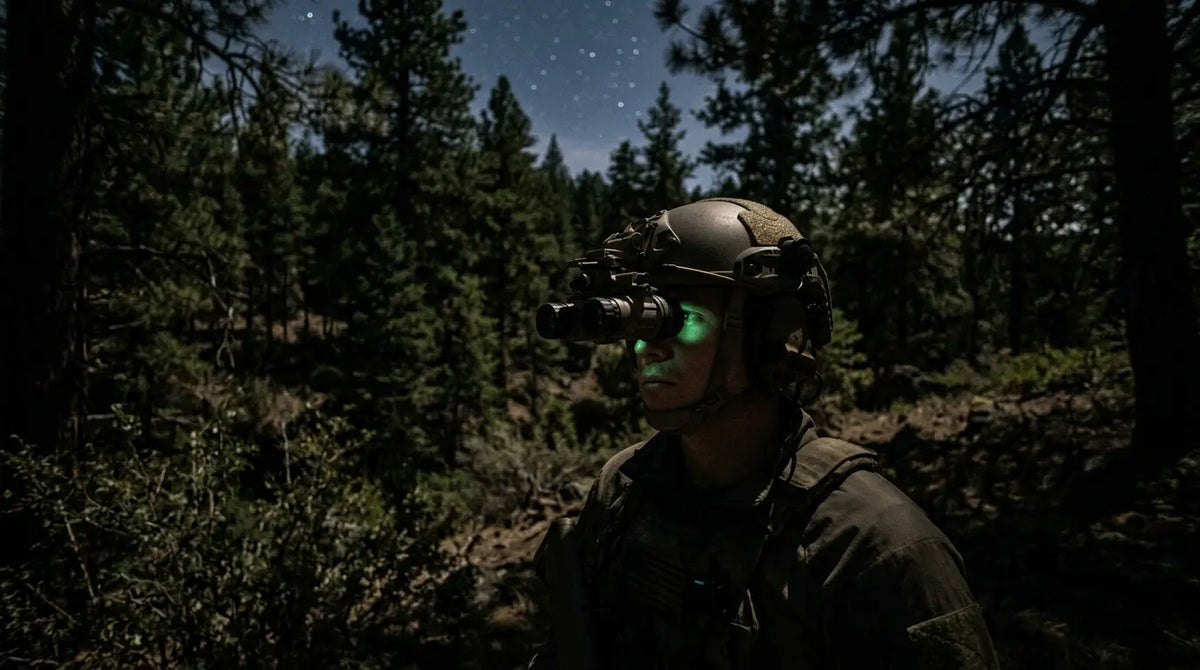
Thermal imaging and image-intensifier night vision both help people observe in low-light settings, but they measure different things. Night vision amplifies or electronically processes available visible and near-infrared light reflected from the scene. Thermal imaging detects infrared radiation associated with object temperature and surface properties. Neither image type is a complete substitute for ordinary vision, and the right tool depends on whether the task is finding a heat contrast or understanding visible scene detail.
| Question | Thermal imaging | Image-intensifier night vision |
|---|---|---|
| What reaches the sensor? | Infrared radiation in the sensor’s designed band. | Available reflected light, commonly visible and near-infrared. |
| Does it need visible illumination? | No; visible darkness alone does not prevent detection of thermal contrast. | It needs usable ambient light or, where supported, supplemental infrared illumination. |
| What does the picture emphasize? | Differences in detected radiation, often associated with surface temperature and emissivity. | Scene shapes and textures represented by reflected light. |
| What can confuse interpretation? | Low thermal contrast, reflective surfaces, atmosphere, obstructions, and processing settings. | Insufficient light, glare, shadows, reflections, and visual clutter. |
| What is a sensible primary role? | Locating heat-signature contrast and scanning when visible light is unavailable. | Reading a more familiar visible-light scene when enough light is available. |
The comparison is about sensing principles, not a universal ranking. A thermal image can make a warm animal or recently operating machine easier to notice against a cooler background, yet give less familiar surface detail than a visible-light image. Night vision can preserve recognizable scene structure but may struggle when illumination is absent or the scene is visually obscured. An operator may use the two approaches for different questions, while following all applicable laws and device guidance.
What affects thermal image quality
Detection is easiest when a target and its background produce a meaningful difference in the radiation reaching the sensor. If both have similar apparent temperatures and surface properties, their boundary may be difficult to distinguish. A subject that was conspicuous earlier can blend into a background after the ground, vegetation, or structures warm or cool. Thermal contrast changes over time and across a scene; it is not a permanent property of the subject.
Weather and intervening material matter too. Rain, fog, humidity, and atmospheric distance can attenuate or scatter infrared energy, reducing contrast or adding path effects. Dense solid objects block the line of sight. Ordinary window glass generally does not transmit the longwave infrared used by many handheld thermal imagers as visible light does, so a camera often shows the glass surface and its reflections rather than a clear thermal view through it. Some thin or sparse vegetation can leave partial openings, but thermal imaging does not see through a solid wall, earth, or dense cover.

Optics and sensor characteristics shape how much scene detail can be represented. Detector resolution is the number of sensing elements, but it is only one part of the system. Lens field of view and focus, distance, target size, motion, atmospheric transmission, display resolution, and image processing all influence the image the viewer perceives. Digital enlargement can make a displayed subject bigger without adding new detector detail. A range claim or ability to identify a target cannot be inferred from resolution alone.
Thermal sensitivity is another distinct concept. A sensitivity figure describes the ability to distinguish small differences in signal under defined test conditions; it is not a guarantee that every real-world target will be clearly visible. Contrast at the target, lens transmission, calibration, processing, and environmental conditions remain important. Likewise, a higher pixel count alone does not guarantee a more useful image in every setting. Compare complete system specifications for the intended task rather than treating any single number as a verdict.
Finally, the display palette changes the way contrast is perceived, not the underlying scene. White-hot and black-hot reverse the light-to-dark mapping; color palettes assign hues to signal ranges. Automatic gain and contrast adjustments can make a changing scene easier to inspect, but they can also alter the apparent relationship between frames. When tracking a subject or comparing observations, understand whether the device is automatically remapping the image and avoid reading color as a universal temperature scale.
A practical way to read a thermal scene
Start by asking what the image actually supports: “There is a detectable thermal contrast in this region” is often safer than “this object is exactly this temperature.” Then consider what might explain the contrast. Is the target warmer or cooler than the background? Could a reflective surface be showing another heat source? Is a pane, rain, fog, or vegetation affecting the path? Has the scene recently changed because of sunlight, wind, or machinery? These questions help separate an observed signal from an assumption about its cause.
- Check the scene context. Compare the target with nearby surfaces rather than interpreting a single bright or dark patch in isolation.
- Account for the surface. Shiny metal and other low-emissivity surfaces may reflect surrounding radiation and produce misleading apparent patterns.
- Respect the line of sight. Glass, walls, ground, and dense barriers do not become transparent because the device is thermal.
- Use the right claim. A contrast image supports detection of a pattern; a precise temperature requires suitable radiometric capability, calibration, and measurement conditions.
- Compare the whole system. Consider lens, field of view, detector, focus, processing, display, and operating conditions together.
These habits are useful whether thermal equipment is used for outdoor observation, wildlife monitoring, equipment inspection, or search work. In professional measurement, follow training and documented procedures. In field observation, treat the display as evidence of relative infrared contrast and seek additional context before making an identification or temperature claim.
Choosing an AGM thermal optic by task
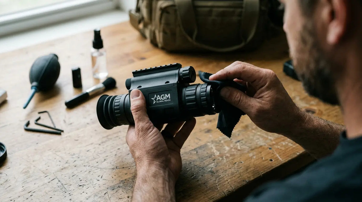
Once the sensing principle is clear, product selection becomes a question of use rather than mystique. A handheld monocular is designed for observation and scanning; a weapon sight or clip-on has a different role and handling requirements. A device page should be consulted for the exact sensor, lens, field of view, controls, and other specifications—those details vary by model and can change. Do not transfer a specification from one product family to another.
For an overview of handheld options, see AGM’s thermal monoculars. That category helps distinguish the form factor intended for handheld scanning from other thermal optics. To understand the complementary role of light-amplifying equipment, AGM also explains thermal optics versus night vision. A reader comparing technologies should match the device to the task: locating thermal contrast, navigating terrain, inspecting a surface, or observing detail are not identical needs.
Before choosing, define the viewing distance and target size, expected background and weather, whether temperature measurement is required, and whether the equipment will be handheld or mounted. Check field of view and focus for the real setting, then review manufacturer documentation for model-specific capabilities and limitations. For use in hunting or other regulated activity, verify current local rules independently. Technology descriptions are not legal advice or a promise of a particular detection outcome.

Frequently asked questions
Can thermal imaging work in total darkness?
Yes. Thermal imagers detect infrared radiation rather than relying on visible light, so darkness by itself does not stop them from forming an image. The scene still needs detectable contrast, and weather, distance, surfaces, and obstructions can affect what appears.
Does thermal imaging show exact temperature?
Not necessarily. Many viewing devices display relative thermal contrast. Reliable temperature measurement requires a radiometric instrument with suitable calibration and corrections for emissivity, reflected radiation, and relevant atmospheric effects.
Can a thermal camera see through glass or walls?
No, not as a general rule. Walls block the line of sight, and ordinary glass often blocks longwave infrared while reflecting thermal energy. The camera may show the barrier’s surface rather than objects behind it.
How is thermal imaging different from night vision?
Thermal imaging senses infrared radiation associated with heat and surface properties. Image-intensifier night vision amplifies available reflected light. Thermal is useful for noticing thermal contrast; night vision provides a more familiar scene view when enough light is available.
Technical sources
- NASA, “Mighty MURI brings the heat to test new longwave infrared radiometer” - uncooled microbolometer principle.
- NASA Landsat Thermal Infrared Sensor (TIRS) - detector, spectral filtering, and thermal-infrared sensing.
- Teledyne FLIR, “How Do Thermal Cameras Work?” -thermal camera components and image formation.
- Teledyne FLIR, Reference Documentation: Thermography - emissivity, reflected radiation, atmosphere, and temperature measurement.
- Usamentiaga et al., “Infrared Thermography for Temperature Measurement and Non-Destructive Testing,” Sensors (2014) - technical review of measurement factors.
- AGM Global Vision thermal monoculars - contextual product category.